Mammals have four kinds of teeth, that differ in shape, function, position in the mouth, and whether or not they are replaced. The four types are incisors, canines, premolars, and molars.
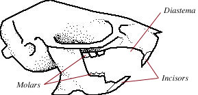
Rats do not have canines (the conical, pointed teeth used for holding prey, defense, and combat), or premolars (grinding teeth behind the canines and in front of the molars). The rats have a long, toothless space in their mouth where the second incisors, canines and premolars would be. This space is called the diastema.
The number of different types of teeth in a species is described with a dental formula, which is written as:
So, the rat's dental formula is:
Anatomy of a tooth
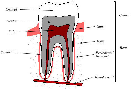
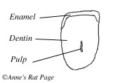
Figure 2. Cross section of a mammalian molar. © anne_rats
Teeth have the same composition as bone. A tooth consists of three layers of mineralized tissues: a hard external layer of enamel forms the crown of the tooth, a hard layer of cementum covers the root. The enamel and cementum surround a layer of softer, living dentin which makes up the bulk of the tooth. Dentin surrounds a soft core of pulp which contains blood vessels and nerves. The periodontal ligament (also called the periodontal membrane) is a fleshy layer that lies between the tooth and the tooth socket. It holds the tooth in place, attaches it to its neighbors, and enables the tooth to resist the stress of chewing (Fig 2).
Rat molars are similar to the molar depicted in Figure 2. Rat incisors, however, have a single, open, root that
continues to grow throughout the rat's life.
Rat incisors
|
|
|
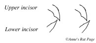
Figure 3. A rat's incisors |
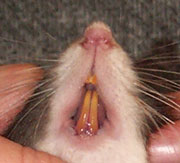
Incisors are specialized for gnawing. The
rat's incisors are open-rooted (Fig 4), which means they grow throughout life (Addison and Appleton 1915). If
allowed to grow without restraint, the rat's incisors would grow in a spiral with an angle of 86º (Herzberg and
Schour 1941). Rats spend their lives gnawing and wearing their teeth down, so the constant growth of their incisors
prevents them from wearing their teeth away.
Rat incisors erupt out of the gum 8-10 days after birth (Addison and Appleton 1915, Schour and Massler 1949). The eruption rate (the rate of growth) of the rat's incisors is very high: the adult rat's upper incisors grow on average about 2.2 mm per week (0.31-0.32 mm per day), and the lower incisors grow about 2.8 mm per week (0.4 mm per day) (Addison and Appleton 1915). It takes about 40-50 days for new tooth generated at the base to reach the tip. The entire tooth is therefore never more than 40-50 days old (Schour and Massler 1942). This rapid growth also keeps the rats' incisors from getting cavities: any cavity would rapidly grow out and be worn away.
The rate of incisor growth varies under different circumstances. If the incisors are trimmed, they grow faster, 1.0 mm per day (+/- 0.1 mm) (Law et al. 2003), so if a rat tends to gnaw on hard substances and thus wear its incisors away quickly, the incisors will grow faster to compensate. Individual incisors can grow at different rates depending on its length and that of its neighbor: if one incisor is shortened, it will grow back faster than the others (Burn-Murdoch 1999). If the upper and lower incisors do not meet properly (called a malocclusion), they cannot be worn away normally and become overgrown.
|
|
|
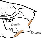
Figure 5. Cross section of a rat's incisor halfway between the gum and the tip of the tooth. Adapted from Addison and Appleton 1915. |
A cross-section of a rat's incisor shows three layers: An inner core of pulp, a surrounding layer of soft dentin, and a layer of hard enamel that covers only the front surface of the tooth (Fig 5, 6). The pulp cavity in the center of the tooth becomes narrower and narrower toward the tip of the incisor (Fig 5). The pulp cavity reaches nearly to the end of the tooth, but at the very end it is filled in with hard material (granular osteodentine), so the sensitive pulp cavity is never actually exposed (Addison and Appleton 1915).
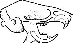
Incisors are kept sharp by gnawing and bruxing, also called thegosis. Because rat incisors have hard enamel only on the front surface, the incisors wear at an angle, with the soft dentin in the back back wearing off before the enamel in the front. This guarantees a sharp, bevel-shaped cutting edge (Fig 6). Rats can actually move their lower jaw so far foward that their lower incisors are in front in front of their upper incisors. When a rat bruxes, its jaw is pulled forward, and its lower incisors sometimes grind behind the upper incisors (wearing down the uppers) and sometimes in front of them (wearing down the lowers) (Fig 7).
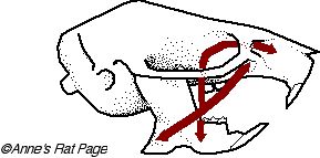
Rats can gnaw powerfully, because the attachment points of the muscles that move the lower jaw up and down are far forward on the nose (Fig 8). This arrangement enables the rat to gnaw very effectively and forcefully. (One of these jaw muscles runs through the eye socket, behind the eyeball. This is why a rat's eyes vibrate in and out, called "eye-boggling," when he bruxes enthusiastically).
When a rat gnaws, its lower jaw shifts forward, bringing the incisors into contact with each other and the molars out of contact with each other. The upper incisors hold the object, and the lower incisors cut against it. Hence, only the incisors are involved in gnawing -- molars do not touch each other when a rat gnaws.
The enamel of rat incisors is hard, harder than iron, platinum and copper. Specifically, measured on Mohs hardness scale, the rat's lower incisors rank 5.5 (diamond is a 10). Human enemel is not quite this hard, measuring 5 on the Mohs hardness scale (ref).
|
|
|
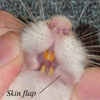
Figure 9. Photo showing skin flap and separation of the rat's lower incisors |
|
|
|
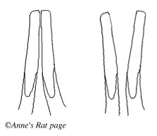
Figure 10. Front of the lower jaw and incisors, showing the incisors closed (left) and open (right). |
The rat's molars are the 12 grinding teeth located at the back of the mouth. They are broad, flat, unpigmented teeth that grind food into a pulp prior to ingestion. When a rat chews, the jaw moves back such that the molars are in contact with each other but the incisors are not. Hence, only the molars are involved in chewing -- the incisors do not touch each other when a rat chews.
Rats have three sets of molars (first, second and third molars). The first molar erupts on the 19th day after birth, the second on the 21st. After the second molar erupts, rats can be weaned. The third molar comes in two weeks later, at around 35-40th day. By age 6 weeks the rat has its complete set of teeth, and by 125 days molar growth slows greatly. After this the molars continue to grow and wear away, but at such a slow rate as to be almost indiscernable (Schour and Massler 1949).
Further reading: