Anatomy of rat and human eyes
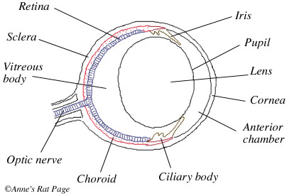
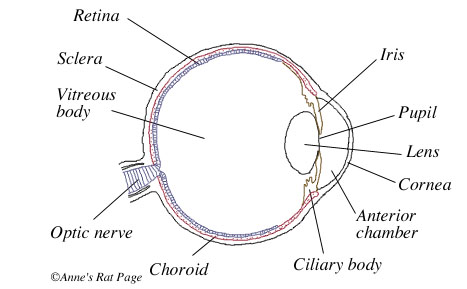
The rat's eye has the same basic structure and function of all mammalian eyes, including the human eye (Figs 1 and 2).
For an interactive, three-dimensional view of the human eye, visit the Anatomy
of the Eye.
When you look at the outside of a rat's eye, what are you seeing? At first glance, rat eyes look like spheres of a single color — little black or pink beads. If you look closely, however, you can distinguish the iris and pupil of a rat's eye. These structures may be easiest to see on a pink-eyed rat (Fig 3).
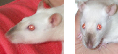
Figure 3. Photograph of a pink-eyed rat, showing the iris and pupil. The pupil is the pale pink circle in the center of the eye, the iris is the darker ring around it. Note that in the photo at right, taken in bright light, the rat has smaller pupils than in the photo at left.
How the eye works: an overview
Here's how the eye works in a nutshell. Light passes through the front structures of the eye — the cornea, lens and so forth. These structures focus the light on the retina, a layer of light receptors at the back of the eye. These receptors translate the image into a neural message which travels to the brain via the optic nerve.
Light passes through a layer of transparent tissue at the front of the eye, called the cornea. The cornea bends the light and is the first element in the eye's focusing system. The light then passes through the anterior chamber, a fluid-filled space just behind the cornea. This fluid is called the aqueous humor, and it is produced by a gland called the ciliary body. The light then passes through the pupil, a round opening in the center of the iris. The iris is a ring of pigmented muscular tissue that controls the size of the pupil, which regulates how much light enters the eye — the pupil grows large in dim light and shrinks to a small hole in bright light. The light passes through the lens, a transparent, biconvex body that helps focus the light from the pupil onto the retina. Light from the lens passes through the vitreous body, a clear jelly-like substance that fills the back part of the eyeball, and is focused onto the retina, a layer of light-sensitive tissue at the back of the eye. The retina contains light-sensitive cells called photoreceptors, which translate the light energy into electrical signals. These electrical signals travel to the brain via the optic nerve. The retina is nourished by the choroid, a highly vascularized membrane that lies just behind the retina. Aside from the transparent cornea at the front of the eye, the eyeball is encased by a tough, white, opaque membrane called the sclera.
Rat vs. Human eyes: a comparison
In both humans and rats, light passes through the cornea, which allows both visible and ultraviolet light (down to 300 nm) to pass through (Hemmingsen and Douglas 1970). Then light passes through the pupil. Like the human pupil, the rat's pupil size is highly variable. Under dim light, the rat's pupil can reach a diameter of 1.2 mm (Block 1969). Under bright light it shrinks to a small opening of about 0.2 mm in diameter (Block 1969, see also Lashley 1932). Pupil diameter changes can be very rapid in the rat: a contraction from 2 mm to 0.5 mm takes only half a second (Lashley 1932).
Next, the light passes through the lens. The lens acts as a filter to block certain wavelengths of light: it is not equally transparent to all colors. Which colors are allowed through differs between species. Human lenses allow only visible light and almost no ultraviolet light to pass through. Rat lenses, on the other hand, allow all visible light plus almost 50% of ultraviolet A light to pass through (Gorgels and van Norren 1992).
The human lens is flexible: the ciliary muscles pull on the lens and thus change its shape. This change in shape causes the light passing through the lens to bend in different ways (called refraction), which allows the lens to focus light on the retina (a process called accomodation). Rats appear unable to change their lenses' shape. For one, rats have a poorly developed ciliary muscle (Lashley 1932, Woolf 1956). For another, relaxing the lens with eyedrops (atropine) does not change its focus (Artal et al. 1998). These findings are consisistent with the idea that rats are unable to change the shape of their lenses, but do not conclusively prove it.
Once the light hits the retina, it is detected by photoreceptors. Humans have two types of photoreceptors: one type that senses light and dark, called rods, and one that senses colors, called cones. We have three types of cones: green, blue, and red. Rats have rods and cones as well, but only two types of cones: green and blue. Therefore, rats are unable to see reds. In addition, the rat's blue cones are sensitive to shorter wavelenghts than our blue cones, which means that rats can see into the ultraviolet (Jacobs et al. 1991). Behavioral experiments have demonstrated that rats can discriminate between greens, blues and ultraviolets, but also that these colors may not have much intrinsic meaning to them (Jacobs et al. 2001). Rats don't have as many cones as we do — 5% of the human retina consists of cones (Hecht 1987), compared to 1% of the rat's retina (LaVail 1976), so their perception of color may be much fainter than ours.
The rat retina has a very "coarse" neural grain. Each neural cell in the rat retina is responsive to a much larger number of photoreceptors than those of the human retina, which increases sensitivity at the expense of acuity. Specifically, the receptive fields of the rat ganglion cells are an order of magnitude larger than those of the human fovea (Brown 1965).
There is great controversy over the refractive state of the rat lens — in other words, whether rats are nearsighted (myopic, Lashley 1932, Weisenfeld & Branchek 1976), farsighted (hyperopic, Block 1969, Massof and Chang 1972), or something in between (e. g. Hughes 1977). However, most agree that rats have poor visual acuity, as demonstrated in behavioral experiments. Rats' acuity is about 20 times worse that that of humans (Wiesenfeld and Branchek 1976, Birch and Jacobs 1979, Artal et al. 1998). Recent experiments by Prusky et al. (2000, 2002) have shown that a normally pigmented rat has about 20/600 vision (1 cpd), and an albino has about 20/1200 vision (0.5 cpd) (see also Birch and Jacobs 1979; Lashley 1930, 1938; Wiesenfeld and Branchek 1976). The rat's poor visual acuity has to do with poor optics combined with the coarse neural grain of the retina (Artal et al. 1998).
As a result of the rat's small eyes and poor visual acuity, rats have an enormous depth of focus (Green et al. 1980). Depth of focus is a property of a visual system that determines the range over which all objects are effectively at the same focal distance. It is determined by the size and acuity of the eye. In humans, the depth of focus of the unaccomodated eye is from 2.3 meters to infinity (Campbell 1957). In rats, the depth of focus is from 7 centimeters to infinity. One consequence of this difference is that humans perceive blurriness after a change of about 1/3 diopter, but rats require a 14 diopter change to perceive any blurriness (Powers and Green 1978).
If you enjoyed this page and would like to read more, visit:
- What do rats see? for more on how rats might see the world
- Rat Cam, for short movies through the eyes of a rat
- Journey into a rat's world for an introduction to rat perception
- The rat's sensory world for articles on the rat's five senses