 By studying rats' eyes and behavior, scientists have a pretty good idea of how a rat sees the world. In
a nutshell, rats are dichromats: they perceive colors rather like a human with red-green colorblindness, but their
color saturation may be quite faint, and color appears to be far less important to them than brightness. Rat vision
is quite blurry, around 20/600 for normally pigmented rats. Albino rats, however, are probably blind or severely
visually impaired, with about 20/1200 vision.
By studying rats' eyes and behavior, scientists have a pretty good idea of how a rat sees the world. In
a nutshell, rats are dichromats: they perceive colors rather like a human with red-green colorblindness, but their
color saturation may be quite faint, and color appears to be far less important to them than brightness. Rat vision
is quite blurry, around 20/600 for normally pigmented rats. Albino rats, however, are probably blind or severely
visually impaired, with about 20/1200 vision.
- What do normally pigmented rats see?
- What do albino rats see?
- Low acuity
- Impaired vision in bright light: dazzling
- Impaired vision in low light
- Few rods, few photoreceptors
- Delayed dark adaptation
- Night blindness
- Problems coordinating what the two eyes see
- Poor depth perception
- Poor motion perception
- Retinal degeneration
- Retinas more easily damaged
- Lens fiber anomalies
- Conclusion
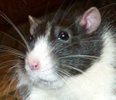
WHAT DO NORMALLY PIGMENTED RATS SEE?
Human and rat retinas have two types of light receptors: cones are sensitive to bright light and color, and rods are sensitive to dim light and cannot see color. Human and rat retinas differ, however, in the types and density of cones in the retina, which has implications for color vision.
Color vision in the retina: Humans have three types of color cones in our retinas. We have "trichromatic" vision, consisting of short-wavelength "blue" cones, middle-wavelength "green" cones and long-wavelength "red" cones.
Rats have just two types of cones (called "dichromatic" vision): a short "blue-UV" and the middle "green" cones (Szel 1992). The "green" cones' peak sensitivity is around 510 nm (Radlwimmer 1998), but the "blue" cones are shifted toward even shorter wavelengths than human blue cones, peaking at 359 nm. This means rats can see into the ultraviolet, they can see colors we can't see (Jacobs et al. 1991; 2001).
About 88% of a rat's cones are the middle "green" type, and 12% are the long blue-UV cones (Jacobs et al. 2001), the blue-UV cones are located in a zone at the bottom of the retina (Szel et al. 1996). For more on how ultraviolet and red-green color vision evolved, see Shi et al. 2001; Yokoyama and Radlwimmer 1999, 2001; Shi and Yokoyama 2003.
Color perception: So, the rat's retina is sensitive to greens and to blue-ultraviolet. Can the rat actually perceive different colors, and distinguish between them? For a long time, rats were throught to be completely colorblind (e.g. Crawford et al. 1990). Recent behavioral experiments, however have shown that rats can indeed perceive ultraviolet light, and with training can distinguish between ultraviolet and visible light, and between different colors in the blue-green range (Jacobs et al. 2001).
What would such vision look like? Animals with red-green colorblindness would be able to distinguish blues from greens, but reds would appear dark to them. They would also have a "neutral" point in the blue-green area of the spectrum: they cannot distinguish these blue-green hues from certain shades of gray. The rat's color vision merges into the ultraviolet, however, so they can see ultraviolet shades that we cannot (see flowers under ultraviolet light to get an idea of what UV looks like).
Rats don't have many cones, though -- 99% of the rat retina consists of rods, which sense only light and dark, and only 1% consists of cones (LaVail 1976), compared to a human's 5% (Hecht 1987). So the rat's perception of color may be fainter than ours, and color cues may not be very important to rats. In fact, brightness appears to be far more important to rats than color. It is easy to train rats to behaviorally differentiate brightnesses, but difficult to train them to behaviorally differentiate colors (Jacobs et al. 2001).
So, while rats are physically capable of distinguishing between ultraviolets, blues, and greens, such differences may not be very meaningful to them. This gets into the whole "just noticeable difference" vs. "just meaningful difference" concept, first introduced by Nelson and Marler (1990).
What is the function of ultraviolet vision? The function of ultraviolet vision in rodents is not well understood yet and is currently an active area of research. Here are some possibilities:
• Urine-mark visibility: Urine is visible under ultraviolet light (humans can see rat urine using a black light). Therefore, when rodents leave urine marks in their environment, these marks may visible as well as smellable (e.g. degus, Chavez et al. 2003; voles, Koivula et al. 1999; mice, Desjardins et al. 1973). Unfortunately, these urine marks may also be visible to predators, such as dirunal raptors. Using ultraviolet cues from the urine marks, kestrels can discriminate between active and abandoned vole trails, thus increasing their hunting success (Viitala et al. 1995).
• The body under UV: different parts of an animal's body may reflect different amounts of ultraviolet light. In degus, for example, the belly reflects more UV light than the back. Therefore, when a degu stands up on its hind legs it exposes its belly to other degus, and ultraviolet vision may come into play. When it stands on all fours its low-reflectance back could help make the degu less visible to predators (Chavez et al. 2003).
• Twilight ultraviolet vision: Ultraviolet light is not available at night, but is abundantly available during the day. Interestingly, there is a significant increase in the ratio of ultraviolet to visible light in the morning and evening twilight hours (Hut et al. 2000). Rats are nocturnal, but they are also active during the twilight hours, starting just before sunset and ending just before sunrise (Robitaille and Bovet 1976). Ultraviolet vision would be advantageous at these twilight times of day. It is therefore possible that ultraviolet sensitivity is retained in rats because it is useful during the twilight hours.
Visual Acuity
The rat's world is very blurry. Visual acuity is measured in cycles per degree (cpd), a measurement of the number of lines that can be seen as distinct within a degree of the visual field. Acuity of humans is about 30 cpd, normally pigmented rats is 1 cpd, and 0.5 for albino rats (Prusky et al. 2002, 2000;also see Birch and Jacobs 1979 who found 1.2 cpd for pigmented rats and 0.34-0.43 cpd for albino rats). If we translate Prusky's cpd measurements into vision chart measurements, a normally pigmented rat has about 20/600 vision, and an albino rat has about 20/1200 vision.
Rat acuity can also be measured by examining the density of ganglion cells in the retina. The denser the ganglion cells, the higher the acuity at that point of the retina. In the rat, the densest area of ganglion cells (defined as the region encompassing 75% of maximum ganglion cell density) is 52.8º wide and is located slightly above and temporal to the optic disk. The maximum density of this area is 6,774 cells/mm2. This isn't very dense -- the densest area of the human retina, the fovea, has up to 38,000 cells/mm2 (Curcio and Allen 1990). The low density of ganglion cells of the rat's retina suggests a maximum visual acuity of 1.5 cpd, which is consistent with the measures found in behavioral acuity experiments (Heffner and Heffner 1992).
Depth of focus: Combined with poor visual acuity, rats have an enormous depth of focus. Depth of focus is the range of distances at which an object is in equivalent focus for an unaccomodated eye. In humans, the depth of focus is from 2.3 meters to infinity (Campbell 1957). In rats, the depth of focus is from 7 centimeters to infinity (Powers and Green 1978), which may be due to the small size of the rat's eye and its poor acuity (Green et al. 1980).
One consequence of this difference in depth of focus is that humans perceive blurriness after a change of about 1/3 diopter, but rats require a 14 diopter change to perceive any blurriness (Powers and Green 1978).
Would tiny eyeglasses help a rat see better?
|
|
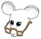
To understand why, imagine that you put lenses of different strengths in front of a human's eyes and a rat's eyes, like the optometrist does when fitting you for glasses. A human can perceive slight differences in the strength of these lenses: a difference as low as 0.3 diopter. A 0.3 diopter lens is a weak lens -- it's weaker than a 0.5 diopter lens, which would correct the vision of a slightly nearsighted person with 20/25 to 20/30 vision.
Therefore, a person with perfect 20/20 vision who puts on 0.3 diopter glasses will detect just a little blurriness. That's the lowest change in lens strength that humans can perceive.
A rat, however, due to its enormous depth of focus, couldn't perceive such a small 0.3 diopter difference in lens strength. So, if you put tiny 0.3 diopter glasses on a rat it would perceive no change in blurriness. It would be the same with 2 diopter glasses, or 6 diopter glasses, or even 10 diopter glasses. In fact, for the rat to perceive any change in blurriness at all, you would need to put thick 14 diopter glasses on it. With such strong lenses the rat would probably perceive a slight increase in blurriness.
The upshot of this is that rat vision is naturally very poor and cannot be corrected with glasses even if you could make glasses that small. Rat eyes are not capable of 20/20 vision. Their eyes' enormous depth of focus, combined with tiny optics, the coarse grain of the retina, their inability to change the shape of their lens to adjust focus, all add up to poor vision. The rat's 20/600 vision is probably as good as it gets. Give a rat immensely strong lenses and its vision would not improve; its vision would become, if anything, slightly blurrier.
- Chart: diopters and 20/something
Orientation
Field of vision vs. binocular depth perception: Rats have eyes on either side of their heads. This position allows for a large field of vision but less binocular vision (Block 1969).
Specifically, the position of the eyes on the head represents a tradeoff between field of vision and binocular vision, which is used in binocular depth perception. Laterally-placed eyes (on either side of the head) scan separate portions of the world, and the field of vision at any one point of time is enormous. Because this position allows the animal to detect threats from many directions at once, it is most common in prey animals (think horses, pigeons, gophers etc.).
Eyes placed on the front of the head have much more overlap. This means a smaller visual field, but more binocular vision. Depth perception is useful for predators who need to coordinate their movements to capture prey, so predators often have forward-facing eyes (think raptors, cats, dogs etc.).
Here's how binocular vision provides depth perception. Each eye sees a slightly different image. The closer an object is, the more different it will look to either eye. The brain processes the disparity between the two images and this provides a sense of depth. Also, focusing on a nearby object makes the eyes converge slightly, and the brain uses information from the eye muscles to calculate how far away the object must be. So, the move overlap between the visual fields of the two eyes, the more binocular depth perception.
Rats don't have as much overlap as we do: their field of binocular vision is only about 76º, while ours is about 105º (Heffner and Heffner 1992). Therefore, rats have a larger field of vision than we do but poorer binocular depth perception.
Depth perception using motion parallax: However, there are many other ways to perceive depth besides binocular vision (e.g. image displacement, motion parallax, and loom). Motion parallax is one of these non-binocular methods of perceiving depth. When one moves one's head from side to side, objects seem to change position relative to each other. Close objects seem to move more than objects that are far away. This is called relative motion parallax, and the brain uses it to calculate relative distance between objects.
An image of the object also moves across the retina. The object's apparent movement across the retina, combined with the amplitude of the head movement are used to calculate absolute distance between the obeserver and the object. This is called retinal or absolute motion parallax (Kral 2003).
Rats use motion parallax to estimate depth. Legg and Lambert (1990) counted the number of vertical head bobs before rats jumped between two platforms. As the gap between the platforms increased, trained rats performed more, and larger head bobs before jumping. They may be adjusting the size of their head movement until they produce a detectable amount of motion parallax (Ellis et al. 1984). As the gap widens, larger head movements are needed. Legg and Lambert (1990) found that that rats could jump accurately even if only the leading edge of the platform was visible, indicating that rats use absolute motion parallax to estimate depth. However, the fact that the rats were reluctant to jump to just a leading edge indicates that they may prefer to use relative motion parallax.
Visual orientation: Rats orient themselves using distant visual cues (Hebb 1938, Lashley 1938). For example, Carr (1913) found that rats trained in a sideless maze lost their way if the maze was rotated, but if the the visual environment was rotated along with the maze their performance didn't suffer (Higginson 1930). The presence of visual cues in mazes speeds up the maze-learning process, while the removal of visual cues causes even a trained rat to make mistakes (Honzik 1936). Rats can discriminate between visual stimuli 51 cm away (Mostafa et al. 2002) and can tell the difference between a blocked and an open maze pathway up to 75 centimeters away (Robinson and Weever 1930).
Rats use their vision for shorter distances, too. For example, rats can jump from a raised platform to another surface over a distance of 30 cm (Lashley 1930).
At very short distances, however, rats may trust their whiskers more than their eyes. In one experiment, rats were placed on a sheet of glass. Half the glass was over a platform, and the other half was over a void. This is called a visual cliff experiment. Animals that rely on visual information to perceive depth, like human children, choose to step onto the glass above the platform instead of the glass over the dropoff. Rats, however, chose the deep and shallow side of the glass in equal amounts: they walked fearlessly on the glass suspended over a dropoff. Their whiskers told them there was a solid surface to walk on. In contrast, rats with clipped whiskers stayed away from the dropoff and chose the shallow side, indicating that without their whiskers they were forced to rely on vision to perceive depth (Schiffman et al. 1970).
Vision damage
Vision and aging: As rats age beyond age two, the rat's retina loses many of its cells, and parts of the retina become enlarged and thickened. The capillaries that feed the retina also become greatly thickened (Weisse 1995). Therefore, old rats probably do not see as well as young rats.
Conclusion
Normally pigmented rats have panoramic, blurry vision with faint greens, blues and ultraviolets. These colors may or may not be meaningful to the rats. This kind of vision probably works just fine for rats: rats can see food and other rats a short distance away, but are still sensitive to incoming hawks or dogs and distant orientation cues.
WHAT DO ALBINO RATS SEE?
Albinos have a number of differences in their visual systems compared to normally pigmented animals. In a nutshell, albinos have no pigment -- no melanin -- in their eyes. This means no pigment in the iris. That's why the iris looks red -- the only color left comes from blood in the capillaries. Albinos also lack pigment deeper in the eye, pigment which normally absorbs light. Without it, light inside the eye scatters. The consequence is that the albino's eye is flooded with light. Over time, the light causes retinal degeneration. In addition to all this, albinos have abnormal neural connections between the eyes and the brain. The end result of all this is that albino rats have very poor vision. Here are the specifics:
Low visual acuity: The albino rat's inability to control levels of incoming light, the scattering of light inside the eye, and gradual retinal degeneration lead to very poor visual acuity. Albino rats have poorer visual acuity than pigmented rats (Prusky et al. 2002), estimated at about 20/1200 vision.
Impaired vision in bright light: dazzling: Albino rats can't control levels incoming light. Normally-pigmented animals have a pigmented iris that surrounds the pupil and controls how much light shines onto the retina. Albinos lack pigment in their irises, so light passes through the iris, dazzling the retina. In bright light, albinos may not see anything at all because their retinas are overwhelmed by the incoming light.
• Few rods, few photoreceptors: Rods require a melanin precursor to develop (dopa). Albinos can't make this. Without it, about 30% of the rat's rods fail to develop (Ilia et al. 2000). Not only does the albino rat have fewer rods, but even early in life the rods it does posess have fewer rod photoreceptors (called rhodopsin) than the rods of pigmented rats (Grant et al. 2001). Rods and their photoreceptors are useful for detecting low levels of light, so albino rats may have trouble seeing in low light conditions.
• Delayed dark adaptation: Albino rats take longer to adapt to the dark than pigmented rats. Specifically, pigmented rats adapt to the dark within 30 minutes, but albino rats take about three hours (Behn et al. 2003). This delay comes from the albino rats' lack of melanin in their eyes. Eyes that lack melanin have reduced bio-availability of calcium (Drager 1985). Calcium plays a key role in a retina's ability to adapt to the low light conditions (called dark adaptation) (Fain et al. 2001).
• Night-blindness in albino rats: conflicting reports: Once dark adaptation is achieved, Balkema (1988) reports that pigmented rats have a much lower dark-adaptation threshold than albinos. In other words, Balkema reports that pigmented rats can see under conditions of much lower light than albino rats. However, Green et al. (1991), Herreros et al. (1992), and Munoz et al. (1994) found no such differences in dark-adaptation thresholds between albino and pigmented rats, indicating no albino night blindness.
Problems coordinating what the two eyes see: There are even further visual differences between albinos and normally pigmented animals, involving the eye-to-brain connection. In normal mammals, the left side of each eye is connected to the right hemisphere of the brain, and the right side of each eye is connected to the left hemisphere. Albinos have a much simpler connection: most of the left eye is connected to the right hemisphere, while most of the right eye is connected to the left hemisphere (Silver and Sapiro 1981). In addition, the deeper neural projections involved in vision are disorganized (Creel et al. 1990). The consequence is that albinos may have trouble coordinating and processing what their two eyes see.
Poor depth perception: The albino rat's poor visual acuity leads to poor visual depth perception. In the visual cliff experiment by Schiffmann et al. (1970) described in the previous section, rats were placed above a sheet of glass over a ledge and dropoff. Pigmented and albino rats with intact whiskers relied on their whiskers instead of their eyes and chose to walk on the glass over the dropoff as often as the glass over the ledge. When the whiskers were clipped, however, the rats were forced to rely on visual cues. Pigmented rats with clipped whiskers chose the glass over the ledge. Most whiskerless albino rats also chose the glass over the ledge, but a large percentage of them (20%-33%) made no choice at all but stood stock still. This failure to choose indicates that albino rats do not use visual information to perceive depth as readily as pigmented rats do. Albinos appear to be more impaired by whisker removal than pigmented rats, probably because their fallback sensory system -- vision -- is so poor.
Pet owners often note that albino rats bob their heads and sway frequently. This bobbing and swaying may be the albino's attempt to increase its perception of depth using its greatly impaired vision.
Poor motion perception: Albino rats have greatly impaired motion perception. They are not motion blind, but they have poor motion perception when compared to pigmented rats. Albino rats require about twice to three times the coherence level to distinguish coherent motion patterns from dynamic noise.
Specifically, Hupfeld and Hoffman (2006) presented rats with moving dot patterns in which dots moved randomly on a screen. A coherent moving pattern was created by a proportion of the dots moving to the right. The percentage of dots moving to the right was called the "percentage of coherence." So, a 100% coherence meant all dots moved to the right; 70% coherence meant 70% the dots moved to the right while 30% moved randomly, and so forth. Both pigmented and albino rats could distinguish between a random pattern and a 100% coherence pattern. When the coherence was reduced, discrimination performance declined in both pigmented and albino rats. Pigmented rats tended to do better, but not significanly so, down to 30% coherence. Below that coherence level pigmented rats did significantly better than albinos. In sum, pigmented rats could discriminate a pattern of 12% coherence from dynamic noise, while albino rats needed about 30% coherence to make the distinction.
Retinal degeneration: In addition to dazzling them, ambient light (even at low intensities) can cause irreversible retinal degeneration (green light; Noell1966). Rods, because they are so sensitive to light, degenerate more easily than cones (Cicerone 1976, Lanum 1978), which reduces the rat's ability to see in low light. Twenty four hours of ambient light is enough to cause some degeneration, and a few weeks is enough to completely degenerate the outer retina (Lanum, 1978), by causing loss of photoreceptors and cell bodies (Wasowicz et al. 2002).
Retinas more easily damaged: When the rat's eyes are subjected to pressure, the albino rat's retina sustains more damage than the pigmented rat's retina (Safa and Osborne 2000).
Lens fiber anomalies: Albino rats also have abnormal lens fibers compared to pigmented rats. When the lens fibers of pigmented and albino rats are observed under the electron microscope, pigmented rat lens fibers have many "ball and socket" joints between them, but albino rats have few of these joints. The membranes of albino rat lens fibres are often ruptured (Yamada et al. 2002).
A note about foveas: Many normally pigmented mammals like humans and other primates have an area of the retina that is packed with cones, called the fovea. The fovea is surrounded by an area with many more rods than cones. The fovea therefore has very sharp, color-sensitive vision that is useful in bright light, while the area surrounding the fovea has blurry, mostly monochromatic vision that is useful in low light conditions. Albinos of these species don't develop a fovea, so they are missing this patch of sharper color vision. However, in rats, even normally-pigmented rats don't develop a fovea (Reese 2002), so fovea hypoplasia in albinos isn't an issue in rat vision.
What if albinos are provided with a missing enzyme during developement? Albinos have a mutated tyrosinase enzyme. This enzyme is essential for the complex chemical reaction leading to melanin. This chemical reaction passes through I-tyrosine, L-DOPA, and I-dopaquinone on its way to melanin. L-DOPA itself plays an important role in retinal development. Lavado et al. (2006) examined transgenic albino mice that had the gene for tyrosine hydroxylase spliced in to their genome. Tyrosine hydroxylase is an enzyme that will do part of what the missing tyrosinase does: it will oxidize I-tyrosine to L-DOPA, but it won't go any further toward melanin. The resulting animals were phenotypically albino, but they did not have many of the visual problems of albinos: they had normal photoreceptors, normal neural pathways in the brain, and improved visual function.
Conclusion
Pigmented rats see a blurry world with only faint color, ranging from green to ultraviolet. The function of such color vision, and whether it is meaningful to rats, is currently unknown.
Pigmented rats rely less on vision and more on smell and hearing than we do. They navigate extensively by whisker touch. Having poor vision is not as great a handicap for the rat as it is for a human. Rats live in a rich world of sound and smell and touch that enables then to navigate effectively in their world.
Albino rats are probably severely visually impaired or blind within a few weeks of opening their eyes. Their retinas degenerate, their brains have trouble coordinating images from their two eyes, and they see poorly in both bright and dim light.
The albino rat will therefore need to compensate for its near-blindnes by using its other senses. Unfortunately, albino rats also appear to have an impaired sense of smell when compared to pigmented rats. However, albinos do appear to have normal hearing.

Human vision

Normally-pigmented rats have blurry dichromatic vision with a little color

Albino rats may see a very blurry, light-dazzled world
For more images of the world through a rat's eyes, visit the Rat Cam page. Visit the rat's eye for more on the anatomy and function of the rat eyes.